Skoltech researchers have investigated the way the composition of small molecules called lipids and metabolites varies in the brain in the first hours and days after death. Looking at samples from humans, mice, and rats, the study considered the levels of nearly 1,000 small molecules. By showing most of them to be fairly stable over the first 48 hours and documenting the transformations undergone by the rest, the work opens the way for further research that could establish the brain-based molecular footprint of schizophrenia, major depressive disorder, and other mental and neurological disorders, whose underlying biology remains poorly understood. This research was published in the journal Biomolecules and was supported by the Russian Science Foundation.
“Consider schizophrenia: We actually don’t know as much about its causes, the affected brain regions, and the molecular changes involved, with obvious repercussions for both diagnosis and treatment,” said the lead author of the study, Marina Zavolskova, a research intern and a PhD student of the Life Sciences program at Skoltech. “To truly understand this and other disorders, people are looking into the differences, say, in protein levels between the healthy and the pathological brain.”
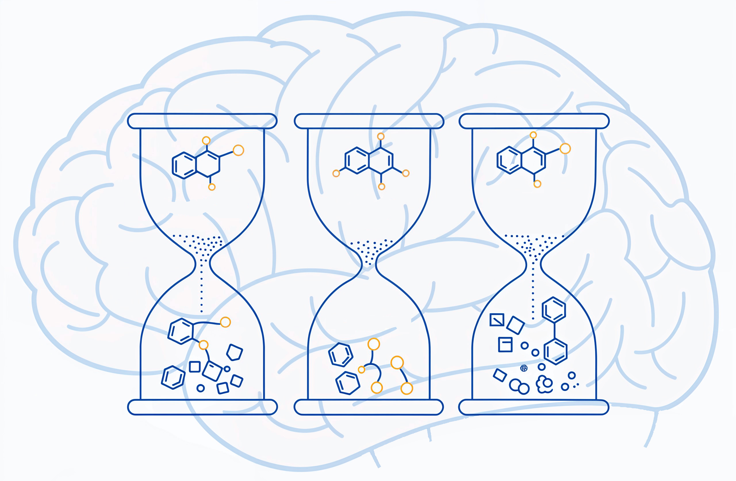
“Lipids and metabolites, too, could be of use,” she goes on. “For example, our colleagues recently reported headway in measuring the lipid levels in the blood of patients with schizophrenia and depression, but analyzing brain tissue is more challenging, because you only get the sample after the death of a patient, and you never know which of the observed effects are due to the disease and which are due to tissue decay. Well, you do know the latter now.”
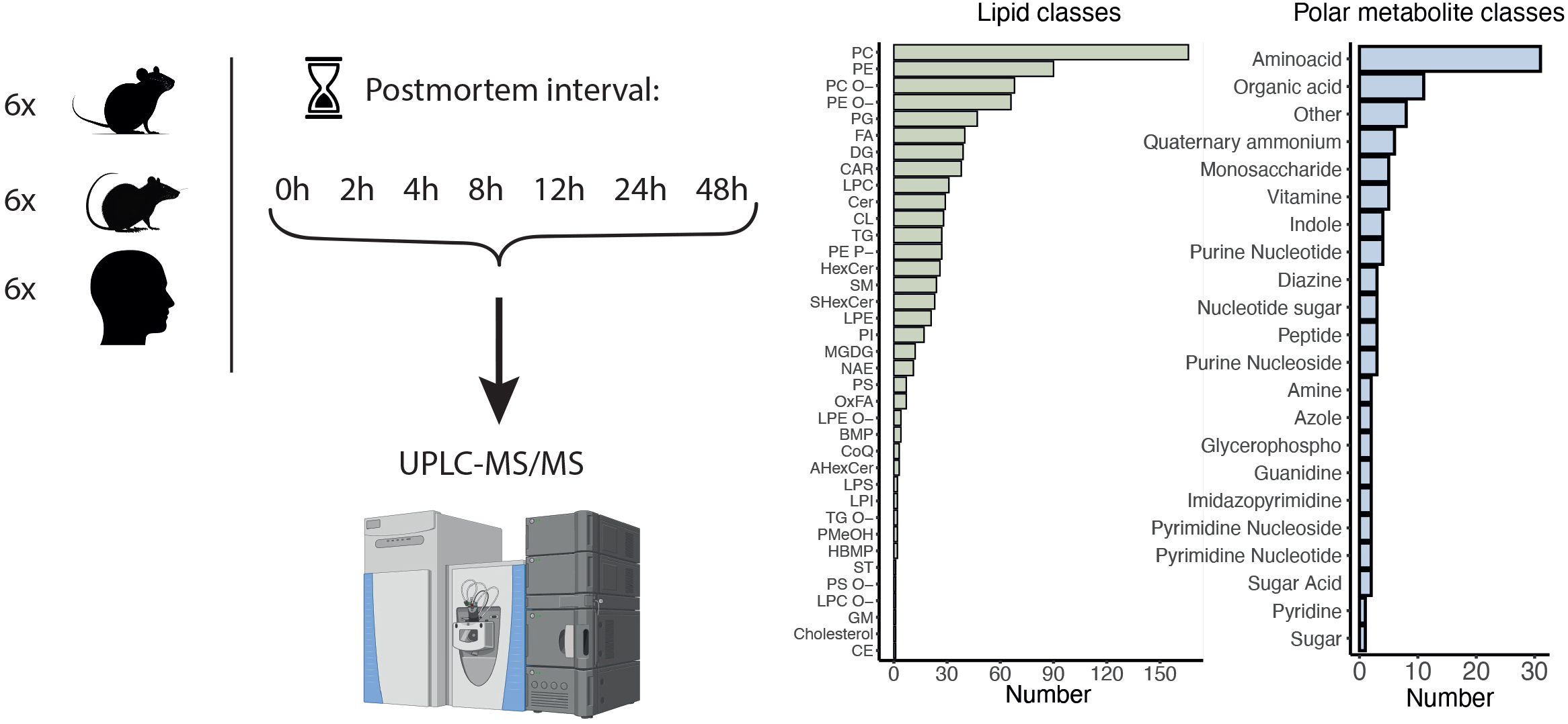
While the study only looks at healthy brains, it establishes a baseline to calibrate the future data from both pathological and healthy brains for their comparison. The team documented the degradation trends for each of the three species, including humans, for 971 small molecules, whose levels were monitored at death, 48 hours later, and five more times in between.