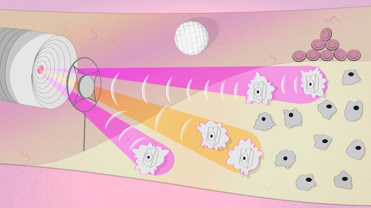
Image. Vascular disco. An artist’s impression of an optoacoustic probe using the technology developed by studies like this. The probe comes into the blood vessel to inspect the composition of an atherosclerotic plaque. The device exposes the constituent cells to light at various wavelengths so they just can’t stand still and picks up the resulting vibes with a mic. Credit: Pavel Odinev/Skoltech
Skoltech researchers and their colleagues have come one step closer to a working optoacoustic endoscopic probe — a device that could slip inside a blood vessel and analyze atherosclerotic plaques by shining laser light on them to make them wobble like a loudspeaker membrane and betray their chemical composition with an ultrasound signature. This could prove useful in robotized microsurgery and medical diagnostics. The study recently came out in ACS Photonics.
Optoacoustic imaging is a promising diagnostic technique that could find applications in routine breast cancer screening, the detection of atherosclerotic plaques or brain lesions, among other things. Unlike a CT scan, optoacoustic sensing does not use X-rays or other harmful radiation but relies on visible light and sound signals — hence the name that combines “optics” and “acoustics.”
Optoacoustics works by exposing biological tissue to pulses of laser light at a wavelength absorbed by some medically significant molecule, say hemoglobin or collagen or perhaps even water. Every pulse heats up the molecules of interest — called biomarkers — causing expansion, followed by contraction in the moments before the next pulse arrives. This periodic oscillation effectively turns the markers into miniature loudspeakers that reveal their locations by emitting ultrasound, which can be picked up by a really sensitive microphone.
The virtues of optoacoustic diagnostics go beyond radiation safety. First, there’s the ability to tune in to specific biomarkers: By varying the wavelength of the laser used to excite acoustic waves, you choose which molecules are targeted. Besides that, ultrasound undergoes fairly low attenuation in biological tissues. That means it can travel farther before the signal dies down, compared to light, affording a deeper look into the body.
“This year the Food and Drug Administration approved an optoacoustic diagnostics system for breast cancer screening. That equipment is not intended to be introduced into the body, though, so both the laser and the microphone are fairly bulky,” the study’s principal investigator, Professor Dmitry Gorin of Skoltech, explains.
“But sometimes a laser just cannot penetrate deep enough, so you have to put a probe inside the body to get a closer look at the insides of a blood vessel or the bladder, for example. This would be useful for inspecting atherosclerotic plaques or possibly even doing microsurgery on them. The probe has to be really thin and should ideally not have any wires,” the researcher adds.
To this end, the researchers are elaborating a setup originally proposed by their English colleagues. The probe is made of an optical fiber that transmits laser pulses and carries a membrane serving as a tiny microphone on its tip. There are two lasers: Pulses from the first one are supposed to arrive at the tip of the probe, pass freely through the membrane, and excite the biomarker. Once it emits acoustic waves, they are picked up by the membrane, and a second laser reads out the signal from the membrane.
“Think of it as a microphone that uses light instead of electricity,” the study’s first author, Nikita Kaydanov, comments. “We used a 100-nanometer film of carbon nanotubes as the microphone membrane. To enable signal readout from the membrane via laser light, we deposited a so-called Bragg mirror of titanium dioxide and silicon dioxide on the nanotube layer. As the mirror oscillates along with the membrane, this modulates the laser signal, enabling readout.”
Another novel feature introduced by the Skoltech researchers was using a hollow-core microstructured waveguide: The optical fiber has an air cavity that runs along its entire length at the center. The benefit of using such fiber is that it can transmit light in the otherwise unavailable mid-infrared range. This can be used to target additional biomarkers: carbohydrates, lipids, and proteins, including those needed to distinguish between atherosclerotic plaques that are relatively harmless and those that require medical attention.
The study takes us one step closer to a working optoacoustic endoscopic probe. Among the team’s findings are data on how the laser heats up the mirror and how this affects its refraction index. This information is essential for correct signal interpretation. “Our experiment also demonstrated signal readout with a laser from the oscillating mirror-coated membrane,” Gorin says. “However, what moved the membrane in our case was not actually the soundwave emitted by biomarkers but the initial laser pulse that lost some of the energy in passing through the membrane.”
According to the researchers, now that they know signal readout works and what the system’s inherent “background” signal is, they can try and pick up actual ultrasound waves from the environment to show the device can function.
Contact information:
Skoltech Communications
+7 (495) 280 14 81
